
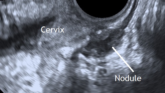
Posterior compartment deep infiltrating endometriotic lesions appear as a hypoechoic thickening of the wall of the bowel or vagina, or as hypoechoic solid nodules which may vary in size and have smooth or irregular contours (Guerreiro et al., 2016).
Bibliography
- Guerriero, S., Condous, G., van den Bosch, T., Valentin, L., Leone, F. P. G., Van Schoubroeck, D., Exacoustos, C., Installé, A. J. F., Martins, W. P., Abrao, M. S., Hudelist, G., Bazot, M., Alcazar, J. L., Gonçalves, M. O., Pascual, M. A., Ajossa, S., Savelli, L., Dunham, R., Reid, S., Menakaya, U., Bourne, T., Ferrero, S., Leon, M., Bignardi, T., Holland, T., Jurkovic, D., Benacerraf, B., Osuga, Y., Somigliana, E. and Timmerman, D. (2016), Systematic approach to sonographic evaluation of the pelvis in women with suspected endometriosis, including terms, definitions and measurements: a consensus opinion from the International Deep Endometriosis Analysis (IDEA) group. Ultrasound Obstet Gynecol, 48: 318–332. doi:10.1002/uog.15955
