
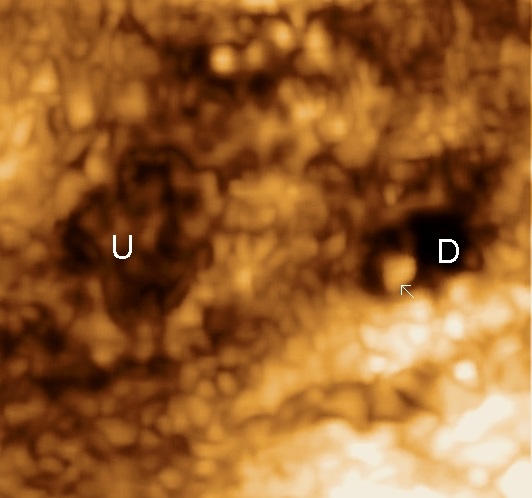
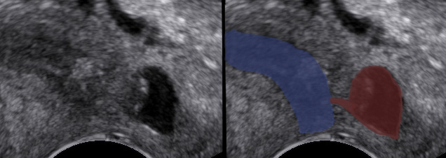
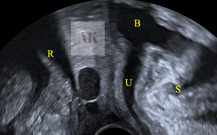
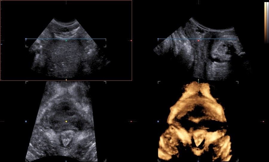
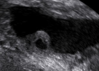
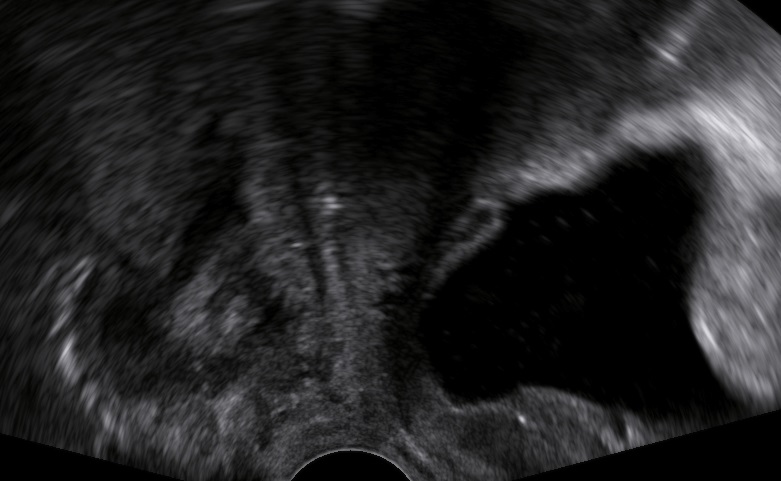
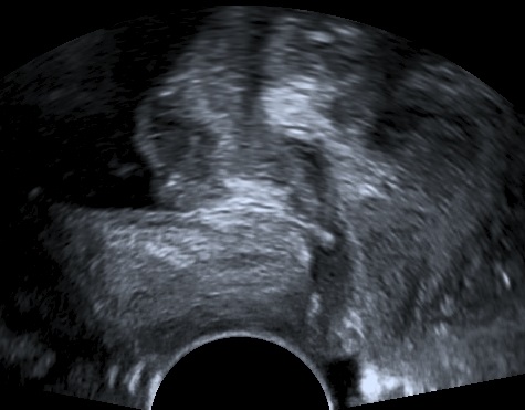
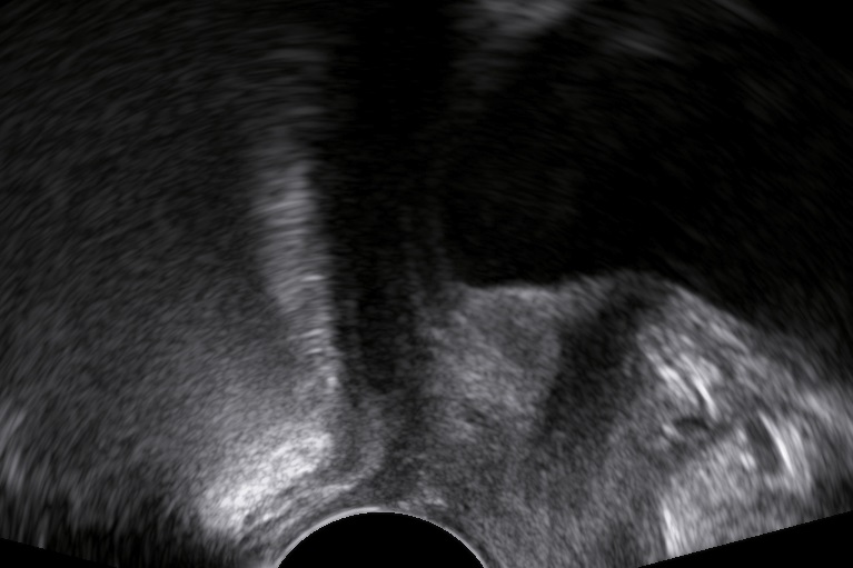
An 80-year-old female patient presents with persisting overactive bladder symptoms. After careful ultrasound examination, a unilocular cystic mass with a single urethral connecting tract containing a calculus was identified as an urethral diverticulum.
Definition
An urethral diverticulum is a rare condition referring to an outpouching of the urethra.
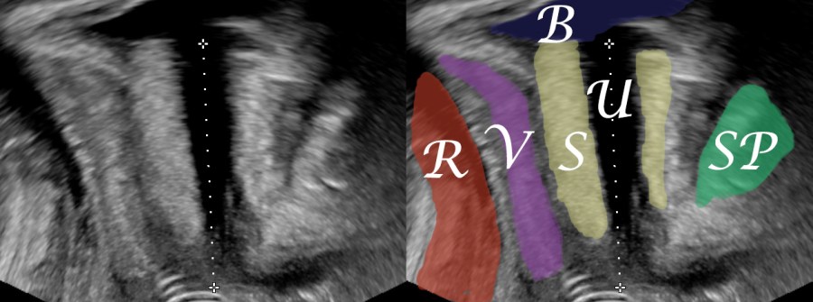
Clinical Image
Diverticula are associated with a multitude of symptoms including urinary incontinence, postmicturition dribble, recurrent urinary tract infections, frequency, urgency, dysuria, dyspareunia, vaginal swelling, anterior vaginal pain or discomfort, urethral discharge and acute urinary retention.
Pathogenesis
Risk factors for its development are recurrent infection of periurethral glands, vaginal birth trauma and previous vaginal or urethral surgery, whereas there have also been a few reports of congenital diverticula.
Diagnosis
The clinician’s suspicion after acquiring a thorough patient’s history should lead to a targetted ultrasound examination combined with digital compression for possible excretion of fluid. The diagnostics may be completed with cystourethroscopy, micturating cystourethrogram, or MRI.
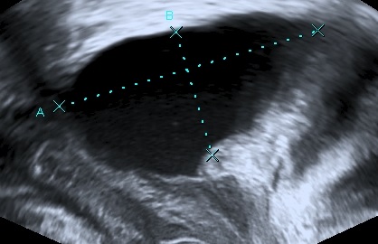
One way to provide a description of a diverticulum is the classification L/N/S/C3 suggested by Leach et. al.:
Location (proximal, mid, distal) / Number of diverticula / Size (diamensions in cm) / Configuration (single, multiloculated or saddle-shaped) / Communication site (proximal / mid / distal) / Continence
Complications
Stone formation, abscess formation and even malignant change.
Management
Transvaginal surgical excision and three-layer closure.
If a pregnant patient presents with an urethral diverticulum consider cesarean section to avoid the risk of fistula.

Classification by Leach et. al:
L (distal) / N (1)/ S (1cm) / C (single) / C (distal) / C (overactive bladder)
You might also find interesting other articles on pelvic floor ultrasound:


Bibliography
- Leach, G. E., Sirls, L. T., Ganabathi, K., & Zimmern, P. E. (1993). L N S C3: a proposed classification system for female urethral diverticula. Neurourology and urodynamics, 12(6), 523–531. https://doi.org/10.1002/nau.1930120602
- Greenwell, T. J., & Spilotros, M. (2015). Urethral diverticula in women. Nature reviews. Urology, 12(12), 671–680. https://doi.org/10.1038/nrurol.2015.230
- Gillor, M., & Dietz, H. P. (2019). Translabial ultrasound imaging of urethral diverticula. Ultrasound in obstetrics & gynecology : the official journal of the International Society of Ultrasound in Obstetrics and Gynecology, 54(4), 552–556. https://doi.org/10.1002/uog.20305
