
Hadlock’s-formula is being widely used for the estimation of fetal weight. Hadlock involved the femur length in his formula and since then it has been an imperative part of every fetal growth assessment.
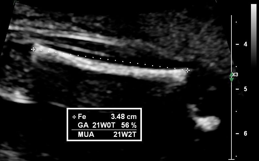
Image quality is of utmost importance in order to perform a correct measurement. Here are some tips on how to achieve this:
- Adjust the image so that bone tissue can be easily identified (usually this means making the image darker).
- Measurement of the femur closest to the probe (this is typically easy to achieve).
- Insonation angle of 90° (to identify the true edges of the femur).
- Appropriate magnification of the femur (at least 30% of the image).
Once the image is satisfactory, the callipers should placed on the outer edges of the femoral bone (outer to outer) excluding the trochanter.
Bibliography
- Hadlock, F. P., Harrist, R. B., Deter, R. L., & Park, S. K. (1982). Fetal femur length as a predictor of menstrual age: sonographically measured. American Journal of Roentgenology, 138(5), 875–878. https://doi.org/10.2214/ajr.138.5.875
- Sarris, I., Ioannou, C., Dighe, M., Mitidieri, A., Oberto, M., Qingqing, W., Shah, J., Sohoni, S., Al Zidjali, W., Hoch, L., Altman, D. G., Papageorghiou, A. T. and for the International Fetal and Newborn Growth Consortium for the 21st Century (INTERGROWTH-21st) (2011), Standardization of fetal ultrasound biometry measurements: improving the quality and consistency of measurements. Ultrasound Obstet Gynecol, 38: 681–687. doi:10.1002/uog.8997
