

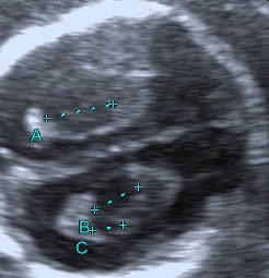
In the second trimester choroid plexus cysts are found in 1–3% of normal fetuses and in 30–50% of trisomy 18 fetuses (Dagklis T. et al., 2008).
The presence of choroid plexus cysts increases the risk for aneuploidy, mainly trisomy 18. In the majority of cases, the final risk will remain small, but will increase as maternal age increases (Chitty L. et al., 1998).
In cases of isolated choroid plexus cysts and low a priory patient’s risk for trisomy 18 we do not recommend invasive testing for fetal karyotyping (Geary M., et al., 1997).

Bibliography
- Dagklis, T., Plasencia, W., Maiz, N., Duarte, L. and Nicolaides, K. H. (2008), Choroid plexus cyst, intracardiac echogenic focus, hyperechogenic bowel and hydronephrosis in screening for trisomy 21 at 11 + 0 to 13 + 6 weeks. Ultrasound Obstet Gynecol, 31: 132–135.
- Chitty, L. S., Chudleigh, P., Wright, E., Campbell, S. and Pembrey, M. (1998), The significance of choroid plexus cysts in an unselected population: results of a multicenter study. Ultrasound Obstet Gynecol, 12: 391–397. doi:10.1046/j.1469-0705.1998.12060391.x
- Geary, M., Patel, S. and Lamont, R. (1997), Isolated choroid plexus cysts and association with fetal aneuploidy in an unselected population. Ultrasound Obstet Gynecol, 10: 171–173. doi:10.1046/j.1469-0705.1997.10030171.x
