
The symptomatic patient typically shows up with one or more of the following (from the most common to the rarest):
- Vaginal bulge or foreign-body sensation
- Chronic constipation
- Vaginal digitation (digital assistance through the vagina for defecation)
- Straining at stool
- Sensation of incomplete bowel emptying
- Faecal incontinence
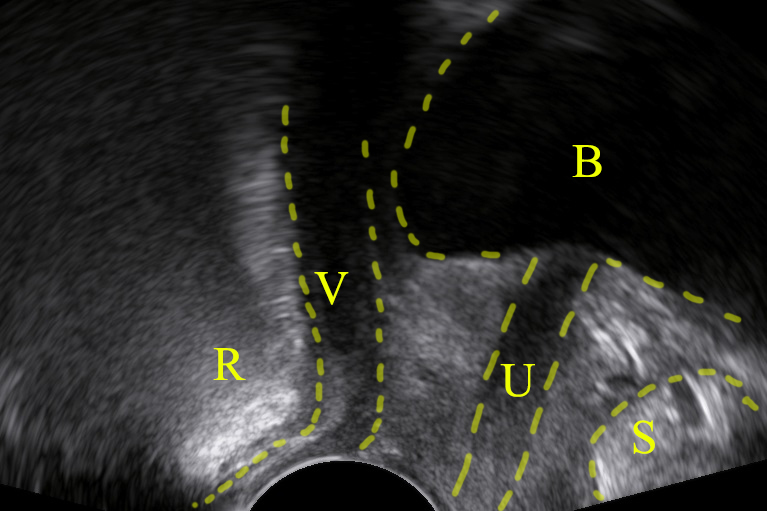
Depicting a rectocele in pelvic floor ultrasound is feasible:

Bibliography
- Dietz, H. P., Zhang, X., Shek, K. L., & Guzman, R. R. (2015). How large does a rectocele have to be to cause symptoms? A 3D/4D ultrasound study. International urogynecology journal, 26(9), 1355–1359. https://doi.org/10.1007/s00192-015-2709-6
