
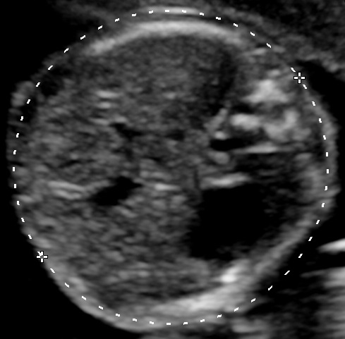
Campbell and Wilkin first described the acquisition of a section orthogonal to the long axis of the fetal body and across the upper abdomen; this was recognized by the typical appearance of the umbilical vein. In cases where the umbilical vein could not be visualized, the fetal stomach was the next most suitable reference point to be used. Then they would take a photograph of the ultrasound image and print it in life size to make their measurements. (Campbell and Wilkin, 1975).
In our days ultrasound machines provide images of much higher quality and measurements can be performed during the examination. However, the defined plane remains the same, as it is easily reproducible. Certainly using today’s technology, a few other anatomical landmarks can also be noted on the same plane.
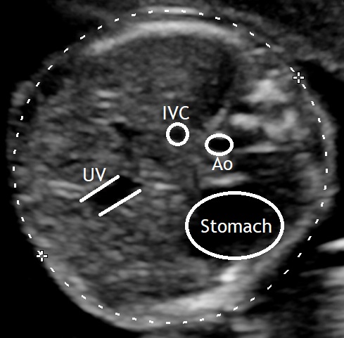
In 1975 Campbell and Wilkin were also the first to describe the prediction of birth weight by ultrasound examination of the fetus. (Smith GC et al, 1997). The Nicolaides group examined numerous models and their results showed that the most accurate prediction of birth weight can be achieved by using the formula devised by Hadlock et al. which involves the measurements of the fetal head and abdominal circumference as well as the femur length (Hammami A, et al, 2018). This underlines the importance of a correct measurement of the abdominal circumference in fetal biometry.
Bibliography
- Smith GC, Smith MF, McNay MB, Fleming JE. The relation between fetal abdominal circumference and birth weight: findings in 3512 pregnancies. Br J Obstet Gynaecol. 1997;104:186–90.
- Hammami A, Zumaeta AM, Syngelaki A, Akolekar R, Nicolaides KH. Ultrasonographic estimation of fetal weight: development of new model and assessment of performance of previous models. Ultrasound Obstet Gynecol 2018; 52: 35–43.
- Campbell, S. and Wilkin, D. (1975), ULTRASONIC MEASUREMENT OF FETAL ABDOMEN CIRCUMFERENCE IN THE ESTIMATION OF FETAL WEIGHT. BJOG: An International Journal of Obstetrics & Gynaecology, 82: 689-697. doi:10.1111/j.1471-0528.1975.tb00708.x
