
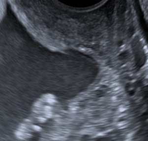
The FMF England presents the following guidelines towards the proper measurement of the cervical length:
- Empty bladder and dorsal lithotomy position.
- No undue pressure on the cervix (longer cervix).
- Median sagittal view of the cervix.
- Calipers between the triangular area of echodensity at the external os and the V-shaped notch at the internal os.
- Examination-time 2-3 minutes (shortest measurement).
The combination of cervical length and obstetric history can be used to predict spontaneous preterm birth. Calculators can be found here: FMF Calculators.

Bibliography
- Celik E, To M, Gajewska K, Smith GC, Nicolaides KH; Fetal Medicine Foundation Second Trimester Screening Group. Cervical length and obstetric history predict spontaneous preterm birth: development and validation of a model to provide individualized risk assessment. Ultrasound Obstet Gynecol 2008;31:549-54.
