

A 30-year-old primigravida in the 16th week of pregnancy shows up with right hypogastric pain, pain at the end of micturition and right flank pain.
Urinalysis showed erythrocytes.
The abdominal ultrasound examination began with an intact pregnancy and a mild maternal hydronephrosis on the right side. The transvaginal sonography showed a distal ureteral calculus with dilatation of the right distal ureter.
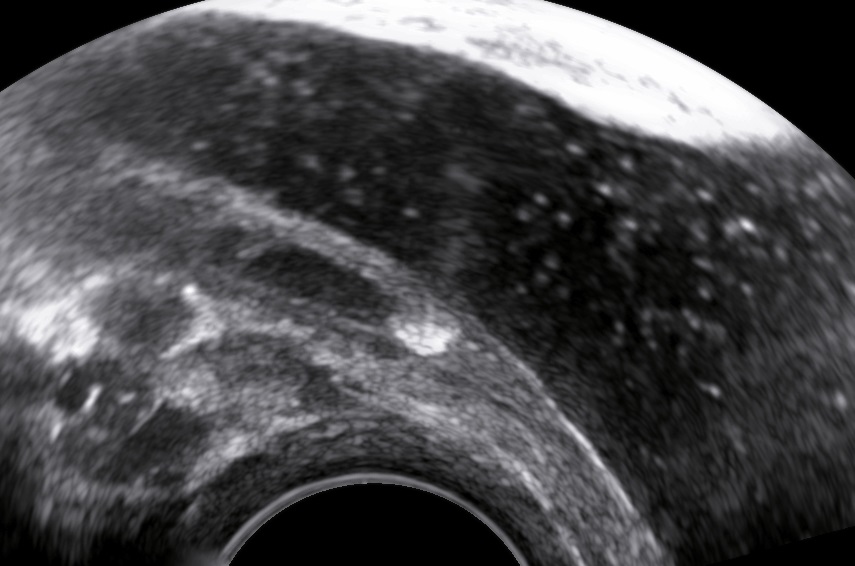
The ureteric stone is perfectly visible with transvaginal sonography. 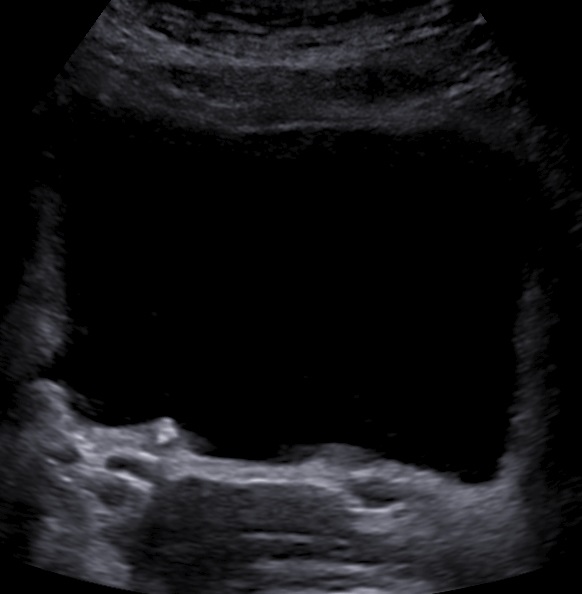
Even abdominal sonography could demonstrate the small ureteral stone. 
A tiny blood clot could be seen inside the bladder. 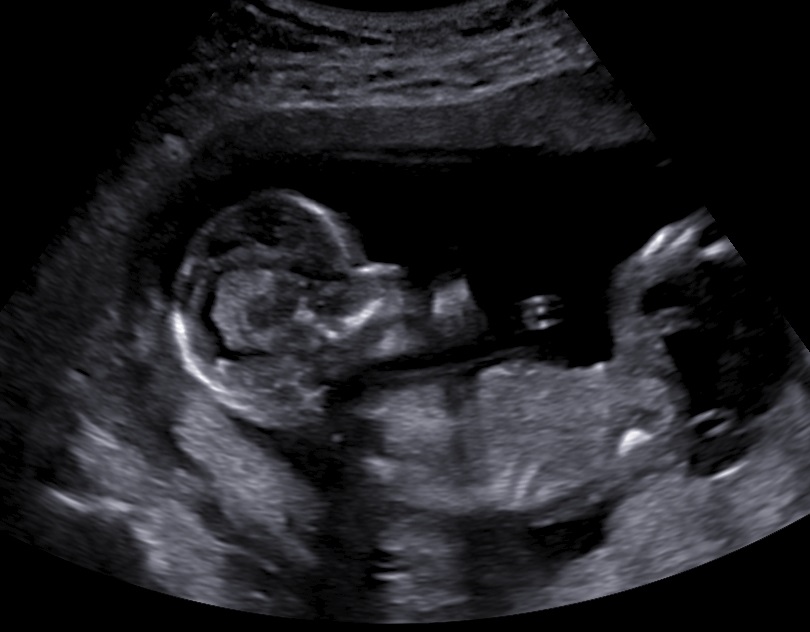
The fetus at 15+0 weeks of pregnancy was normal.
The stone only measured 3.4mm and after conservative treatment (analgetics and hydration) the patient managed to pass it.
In conclusion, transvaginal sonography provides a perfect way to evaulate distal ureteral calculi and completely avoid radiation, especially in pregnancy.
Bibliography
- Laing, F. C., Benson, C. B., DiSalvo, D. N., Brown, D. L., Frates, M. C., & Loughlin, K. R. (1994). Distal ureteral calculi: detection with vaginal US. Radiology, 192(2), 545–548. https://doi.org/10.1148/radiology.192.2.8029429
