
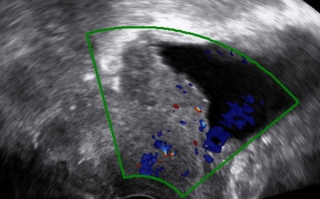
Transvaginal as well as transrectal ultrasound can and should be used in the preoperative work-up of cervical cancer.
It can evaluate the exact location of the tumor, its growth pattern, its size, the extent of invasion and provides the advantage of the dynamic pelvic examination.
Topography/growth pattern
- Exocervix/exophytic tumor growth,
- Endocervix/infiltrative tumor growth
Tumor size
- Craniocaudal diameter in sagittal plane
- Anteroposterior diameter in sagittal plane
- Laterolateral diameter in transverse plane
- For fertility-sparing procedures is the tumor at least 1cm away from the internal cervical os?
Investigate Invasion
a. Partial stromal invasion (tumor – pericervical fascia distance)
b. Complete stromal invasion
c. Parametrial invasion (tumor invades the pericervical fascia laterally)
d. Infiltration of the vesicovaginal septum (tumor invades the pericervical tissue anteriorly)
e. Infiltration of the rectovaginal septum (tumor invades the pericervical tissue posteriorly)
f. Tumor extends to the vagina (fornices are obliterated)

Bibliography
- Testa, A.C., Ludovisi, M., Manfredi, R., Zannoni, G., Gui, B., Basso, D., Di Legge, A., Licameli, A., Di Bidino, R., Scambia, G. and Ferrandina, G. (2009), Transvaginal ultrasonography and magnetic resonance imaging for assessment of presence, size and extent of invasive cervical cancer. Ultrasound Obstet Gynecol, 34: 335-344. doi:10.1002/uog.7325
- Pinkavova, I., Fischerova, D., Zikan, M., Burgetova, A., Slama, J., Svarovsky, J., Dundr, P., Dusek, L. and Cibula, D. (2013), Transrectal ultrasound and magnetic resonance imaging in the evaluation of tumor size following neoadjuvant chemotherapy for locally advanced cervical cancer. Ultrasound Obstet Gynecol, 42: 705-712. doi:10.1002/uog.12455
- Fischerová, D., Cibula, D. (2019), The role of ultrasound in primary workup of cervical cancer staging (ESGO, ESTRO, ESP cervical cancer guidelines), Winter;84(1):40-48.
- Huang, W.‐C., Yang, J.‐M., Yang, Y.‐C. and Yang, S.‐H. (2006), Ultrasonographic characteristics and cystoscopic correlates of bladder wall invasion by endophytic cervical cancer. Ultrasound Obstet Gynecol, 27: 680-686. doi:10.1002/uog.2775
- Fischerova, D. (2011), Ultrasound scanning of the pelvis and abdomen for staging of gynecological tumors: a review. Ultrasound Obstet Gynecol, 38: 246-266. doi:10.1002/uog.10054
- Gaurilcikas, A., Vaitkiene, D., Cizauskas, A., Inciura, A., Svedas, E., Maciuleviciene, R., Di Legge, A., Ferrandina, G., Testa, A.C. and Valentin, L. (2011), Early‐stage cervical cancer: agreement between ultrasound and histopathological findings with regard to tumor size and extent of local disease. Ultrasound Obstet Gynecol, 38: 707-715. doi:10.1002/uog.9037
