

The nuchal translucency (NT) measurement is an integral part of the first-trimester screening for fetal anomalies. The following prerequisites are important for a proper measurement:
- Operator with specialized training and continuing certification (www.fetalmedicine.com)
- Scan between 11 and 13+6 weeks corresponding to a CRL measurement of between 45 and 84 mm.
- Fetus in a neutral position.
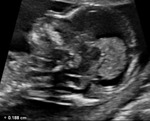
- Magnification of the image including only the fetal head and upper thorax.
- The amniotic membrane should be identified separately from the fetus.
- Medial view of the fetal face in a true sagittal section of the fetus, defined by the presence of the echogenic tip of the nose and rectangular shape of the palate anteriorly, the translucent diencephalon in the center and the nuchal membrane posteriorly.
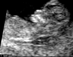
- Ultrasound machine precision of 0.1 mm.
- Calipers should be placed on-on to measure NT as the maximum distance between the nuchal membrane and the edge of the soft tissue overlying the cervical spine.
- Use of the maximum measurement in case two or more images meet all the criteria.
- Multiple pregnancy requires special considerations, taking into account chorionicity.
Optimizing the ultrasound image is of paramount importance to get a good NT. This is exactly why you might find this next article interesting:

Bibliography
- (2013), ISUOG Practice Guidelines: performance of first-trimester fetal ultrasound scan. Ultrasound Obstet Gynecol, 41: 102–113. doi:10.1002/uog.12342
