
The female urethral length varies from 19 to 45 mm (Promian A et al, 2018). Anatomically it can be divided into 5 parts of equal length. The proximal part (1/5) is surrounded by the structures forming the bladder neck. The next two parts (2/5 and 3/5) are surrounded by the striated M. sphincter urethrae and smooth muscle tissue. Around the fourth part (4/5) appear two other muscles forming a bow in front and along the sides of the urethra, the M. compressor urethrae and the M. sphincter urethrovaginalis. The distal part (5/5) is not surrounded by muscular tissue (Perucchini D et al, 2009).
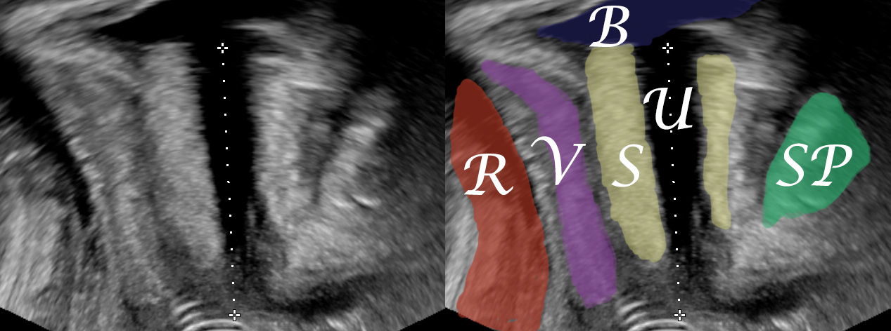
A median sagittal image of the urethra should be achieved with the ultrasound probe placed on the external urethral meatus. The pubic symphysis should be on the right as a reference point. Care should be taken that no pressure is applied on the urethra, something that would lead to a measurement shorter than the true urethral length. Thus there should be adequate distance between the ultrasound probe and the urethral sphincter complex. The urethral length is measured along the anechoic urethra (Wlazlak E et al, 2016).

Bibliography
- Perucchini, Daniele, et al. “4. Funktionelle Beckenbodenanatomie.” Urogynäkologie In Praxis Und Klinik, 2nd ed., De Gruyter, 2009, pp. 25–28.
- Wlazlak E, Kociszewski J, Suzin J, Dresler M, Surkont G. Urethral length measurement in women during sonographic urethrocystography – an analysis of repeatability and reproducibility. Journal of Ultrasonography. 2016;16(64):25-31. doi:10.15557/JoU.2016.0003.
- Pomian A, Majkusiak W, Kociszewski J, et al. Demographic features of female urethra length. Neurourology and Urodynamics. 2018;1–6. https://doi.org/10.1002/nau.2350
