
Adenomyosis is characterized by the presence of ectopic endometrial glands and/or stroma within the myometrium. The definitive diagnosis is made through histological evaluation of the surgical specimen (Andres M. P., et al, 2018).
In order to describe and document the ultrasonographic suspicion of adenomyosis, a classification and reporting system was recently published:
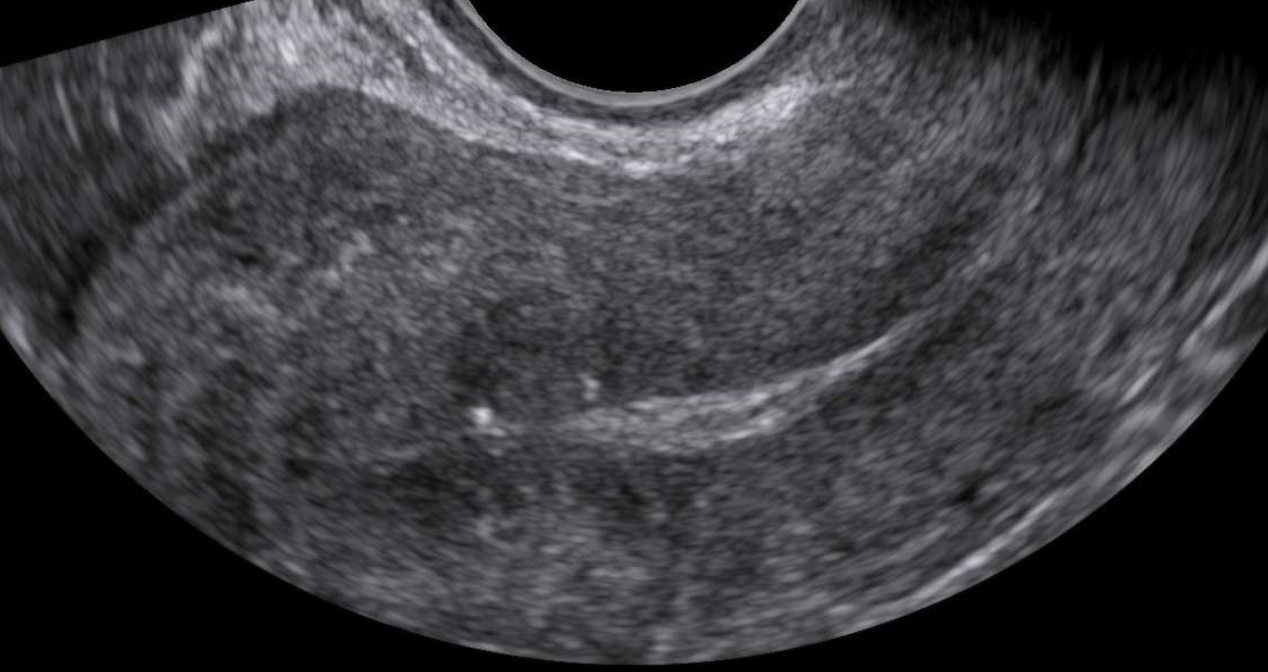
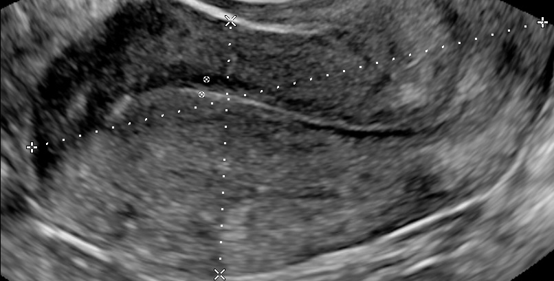
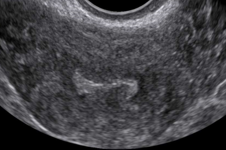
- Characteristics associated with adenomyosis according to the MUSA criteria (enlarged globular uterus, asymmetrical thickening of the myometrium, myometrial cysts, echogenic subendometrial lines and buds, hyperechogenic islands, fan shaped shadowing, an irregular or interrupted junctional zone, and translesional vascularity on color Doppler)
- Location (anterior, posterior, lateral left, lateral right or fundal)
- Focal, diffuse, mixed-type, or adenomyoma (focal adenomyosis refers to cases where less than 25% of the myometrium of the corpus uteri is involved and more than 25% of the circumference of the lesion is surrounded by normal myometrium, estimated on that sagittal uterine section where the adenomyotic lesion appears the largest. When focal adenomyosis is distinctly demarcated and surrounded by hypertrophic myometrium, it is called an adenomyoma.)
- Cystic or non-cystic (cysts at least 2mm)
- Myometrial layer involvement (type 1: the junctional zone, type 2: the middle myometrium, type 3: the outer myometrium – the layer between the serosa and the vascular arcade)
- Disease extent (mild: <25% of uterine volume, moderate: 25-50% or severe: >50%)
- Lesion size
It sounds sensible that the examiner records cycle day and/or current hormonal use (Van den Bosch, T., et al, 2019).
Lastly, elastography appears promising in facilitating the diagnosis of adenomyosis, however more research is needed (Frank M., et al, 2015).

Bibliography
- Andres, M. P., Borrelli, G. M., Ribeiro, J., Baracat, E. C., Abrão, M. S., & Kho, R. M. (2018). Transvaginal Ultrasound for the Diagnosis of Adenomyosis: Systematic Review and Meta-Analysis. Journal of Minimally Invasive Gynecology, 25(2), 257–264. https://doi.org/10.1016/j.jmig.2017.08.653
- Van den Bosch, T. , de Bruijn, A. M., de Leeuw, R. A., Dueholm, M. , Exacoustos, C. , Valentin, L. , Bourne, T. , Timmerman, D. and Huirne, J. A. (2019), Sonographic classification and reporting system for diagnosing adenomyosis. Ultrasound Obstet Gynecol, 53: 576-582. doi:10.1002/uog.19096
- Frank, M., Schäfer, S., Möllers, M., Falkenberg, M., Braun, J., Möllmann, U., … Schmitz, R. (2015). Importance of Transvaginal Elastography in the Diagnosis of Uterine Fibroids and Adenomyosis. Ultraschall in Der Medizin – European Journal of Ultrasound, 37(04), 373–378. doi:10.1055/s-0035-1553266
