
The Hadlock-formula is being widely used for the estimation of fetal weight. Hadlock explained the reasons behind the choice of the plane section for sonographic measurement of the bi-parieral diameter (BPD).
- The fetal head should occupy at least 30% of the image and should appear as an ovoid structure, while the midline echo or falx cerebri should be vertical to the ultrasound beam.
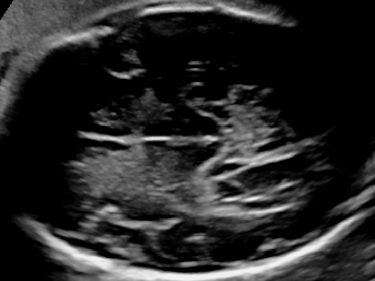
- Acquire a symmetrical axial plane of section with the widest transverse and frontal-occipital diamensions of the fetal head where the falx cerebri, the cavum septi pellucidi, both thalami and both cerebral peduncles are visible (Sarris et al, 2011).
- Place the calipers at the outer surface of the skull side near the transducer and at the inner margin of the opposite skull side. While outer-to-outer measurements have also been described, we suggest the use of the outer-inner method because this was the method used by Hadlock (Hadlock et al, 1982).

Bibliography
- Sarris, I., Ioannou, C., Dighe, M., Mitidieri, A., Oberto, M., Qingqing, W., Shah, J., Sohoni, S., Al Zidjali, W., Hoch, L., Altman, D. G., Papageorghiou, A. T. and for the International Fetal and Newborn Growth Consortium for the 21st Century (INTERGROWTH-21st) (2011), Standardization of fetal ultrasound biometry measurements: improving the quality and consistency of measurements. Ultrasound Obstet Gynecol, 38: 681–687. doi:10.1002/uog.8997
- Hadlock FP, Deter RL, Harrist RB, et al. Fetal biparietal diameter: Rational choice of plane section for sonographic measurement. AJR Am J Roentgenol. 1982;138:871-874.
