
Imaging of the ureters is feasible and trained gynecologists should be able to perform it when needed (Aas-Eng, K., et al, 2019).
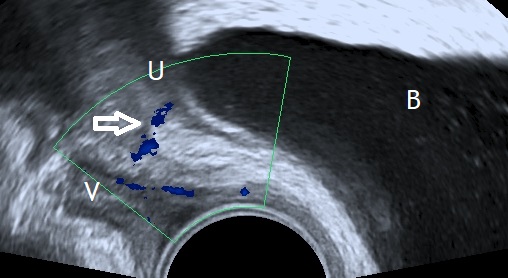
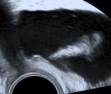
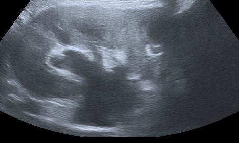
The following images represent an abnormal passageway between the ureter and the vaginal apex following a complicated hysterectomy.

Bibliography
- Aas‐Eng, K. , Salama, M. , Sevelda, U. , Ruesch, C. , Nemeth, Z. and Hudelist, G. (2019), Learning curve for detection of the distal part of ureters by transvaginal sonography (TVS): a feasibility study. Ultrasound Obstet Gynecol. Accepted Author Manuscript. doi:10.1002/uog.20379
