

There is a general tendency for myomas to grow in size during pregnancy (De Vivo, A. et al., 2011). Obstetrical complications related to myomas include abdominal pain, preterm delivery, high rate of cesarean section, intrauterine fetal death and postpartum hemorrhage (Lee, Y., et al., 2012). Specifically, the presence of multiple myomas is associated with a heavier blood loss at delivery (Andreani, M., et al., 2009) and only the myomas that are located in the lower uterine segment are associated with a higher rate of cesarean section (Tajima, A. and Yoshida, K., 2014).
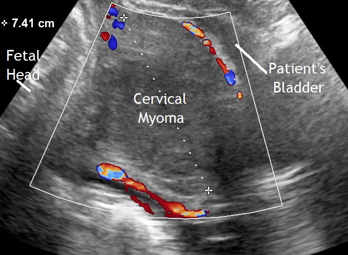
Our first image shows a cercical myoma with a diameter of 7 cm. Cesarean section was performed.
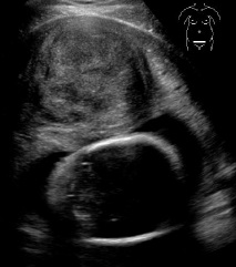
Our second image shows a myoma at the level of the uterine isthmus. On vaginal examination the fetal head could not be palpated. Here was a cesarean delivery performed with increased intraoperative bleeding.
The next patient had at least five myomas with increasing size during pregnancy.


The following clip shows a cervical myoma in the third trimester documented through a cranial movement of the transversally placed abdominal transducer.
The last video shows a subserous myoma in the area of the uterine fundus without complications throughout pregnancy and delivery.
Bibliography
- De Vivo, A., Mancuso, A., Giacobbe, A., Maggio Savasta, L., De Dominici, R., Dugo, N., Dugo, C. and Vaiarelli, A. (2011), Uterine myomas during pregnancy: a longitudinal sonographic study. Ultrasound Obstet Gynecol, 37: 361–365. doi:10.1002/uog.8826.
- Lee, Y., Park, J., Jung, S., Kwon, J., Kim, Y., Park, Y. and Son, G. (2012), P20.01: The influence of large uterine myoma on pregnancy outcome. Ultrasound Obstet Gynecol, 40: 249. doi:10.1002/uog.12037.
- Andreani, M., Vergani, P., Ghidini, A., Locatelli, A., Ornaghi, S. and Pezzullo, J. C. (2009), Are ultrasonographic myoma characteristics associated with blood loss at delivery?. Ultrasound Obstet Gynecol, 34: 322–325. doi:10.1002/uog.7319.
- Tajima, A. and Yoshida, K. (2014), P17.15: The relation of myoma located in the lower uterine segment and the high risk of Caesarean section. Ultrasound Obstet Gynecol, 44: 289–290. doi:10.1002/uog.14348.
