
Definition
A congenital dilation of the distal portion of the ureter, frequently but not exclusively associated with ureteral duplication.
Clinical Image & Complications
Large ureteroceles can cause acute urinary obstruction, hydronephrosis, painful vaginal mass, hematuria, dysuria and urinary tract infections. A chronic obstruction may lead to loss of kidney function.

Pathogenesis
Abnormal development between the accessory ureteral bud and urogenital sinus or a defect in the muscular wall of the ureter.
Diagnosis
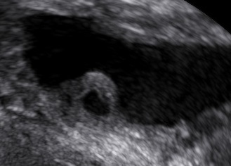
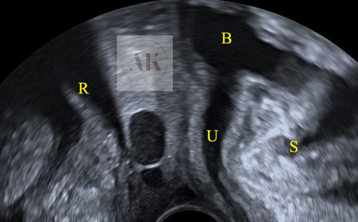
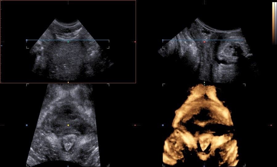
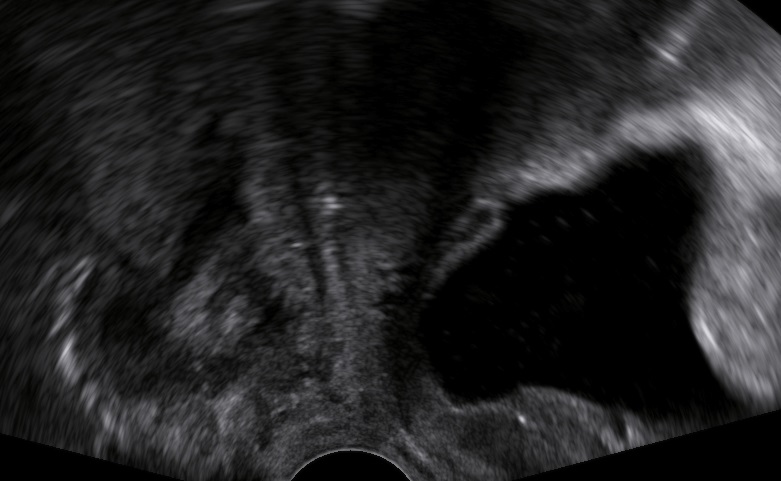
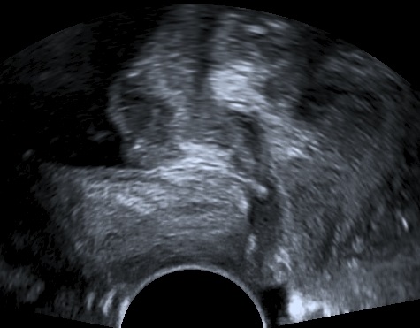
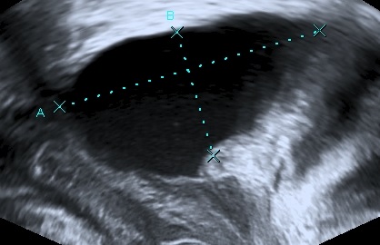
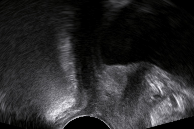
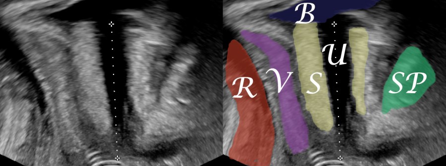
On ultrasound the ureterocele within the bladder appears like a cyst within a cyst. During live examination the ureterocele can be seen to inflate and deflate.
Traditionally the diagnosis of an ureterocele was made with an intravenous pyelogram (cobra head sign).
Management
Usually conservative follow-up. Transurethral resection in case of serious symptoms.
You might also find interesting other articles on pelvic floor ultrasound:


Bibliography
- Tam, T., Pauls, R.N. Embryology of the urogenital tract; a practical overview for urogynecologic surgeons. Int Urogynecol J 32, 239–247 (2021). https://doi.org/10.1007/s00192-020-04587-9
