
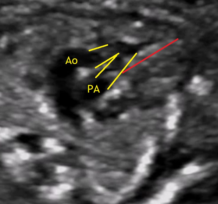
There are four types of ventriculo-arterial connection: (1) concordant, (2) discordant, (3) double-outlet and (4) single-outlet. Double-outlet is when more than 50% of each great artery is connected to the same ventricular chamber. Double-outlet can occur from a morphologically right, a morphologically left or from a ventricle of indeterminate morphology (Carvalho J., et al., 2005).
One out of 10.000 children is born with a double outlet right ventricle (DORV), which means both great vessels arise either entirely or predominantly from the right ventricle. The aorta can be found in right anterior (D-transposition type DORV), right posterior (tetralogy of Fallot type DORV), right lateral (side by side) or left anterior position (L-transposition type DORV). Commonly there is also a ventricular septal defect (VSD) which can be subaortic, subpulmonary, subaortic and subpulmonary (doubly commited) and remote (nonrelated to both arteries) (Abuhamad A., Chaoui R., 2010).
DORV is found in fetuses with a large spectrum of associated cardiac and extracardiac lesions. The overall prognosis for fetuses with DORV is poor, not only related to the primary lesion but also depending on the associated abnormalities (Gedikbasi A. et al., 2007).
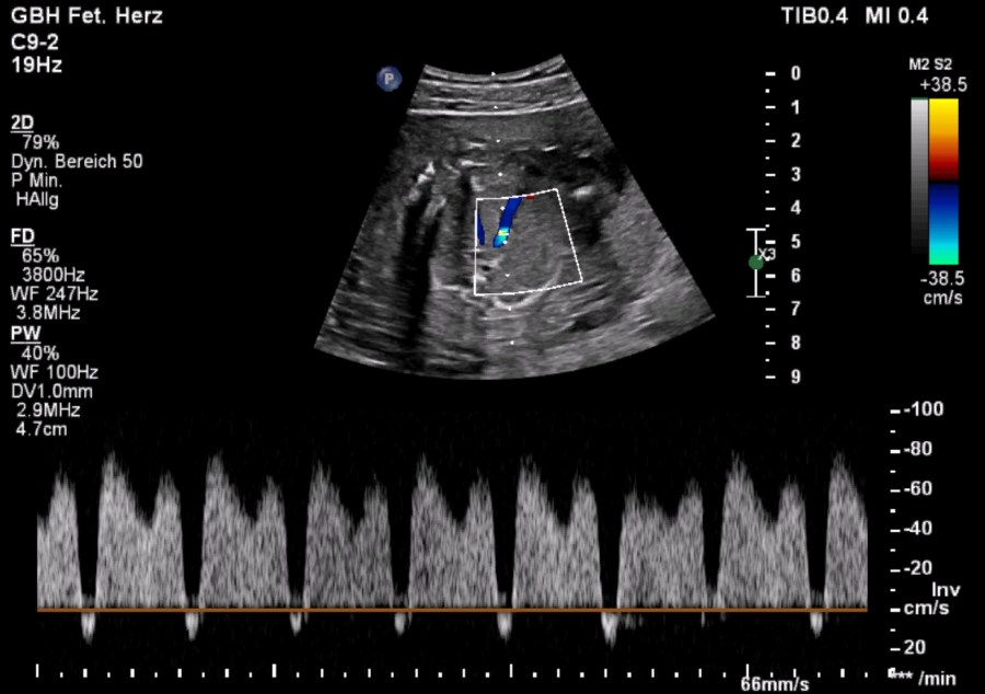
Our featured video shows a case of DORV diagnosed in the 19th week of pregnancy. The image below shows the pathological doppler findings of the ductus venosus.
Bibliography
- Carvalho, J. S., Ho, S. Y. and Shinebourne, E. A. (2005), Sequential segmental analysis in complex fetal cardiac abnormalities: a logical approach to diagnosis. Ultrasound Obstet Gynecol, 26: 105–111. doi:10.1002/uog.1970
- A practical guide to fetal echocardiography, second edition, Alfred Abuhamad, Rabih Chaoui, 2010.
- Gedikbasi, A. G., Oztarhan, K. O., Gul, A. G. and Ceylan, Y. C. (2007), P39.08: Double-outlet right ventricle: prenatal diagnosis and fetal outcome. Ultrasound Obstet Gynecol, 30: 598–599. doi:10.1002/uog.4882
