
Diagnosing a vesicovaginal fistula depends a lot on the patient’s history, typically involving absolute urinary incontinence during the day and during the night since the surgical intervention.
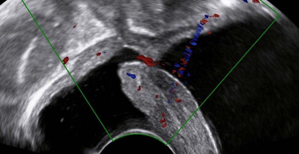
The following images and videos represent an abnormal passageway between the bladder and the vaginal apex following a complicated hysterectomy.
Bibliography
- Volkmer, B. G., Kuefer, R., Nesslauer, T., Loeffler, M., & Gottfried, H. W. (2000). Colour Doppler ultrasound in vesicovaginal fistulas. Ultrasound in medicine & biology, 26(5), 771–775. https://doi.org/10.1016/s0301-5629(00)00210-6
- Sohail, S., & Siddiqui, K. J. (2005). Trans-vaginal sonographic evaluation of vesicovaginal fistula. JPMA. The Journal of the Pakistan Medical Association, 55(7), 292–294.
- Stamatakos, M., Sargedi, C., Stasinou, T., & Kontzoglou, K. (2014). Vesicovaginal fistula: diagnosis and management. The Indian journal of surgery, 76(2), 131–136. https://doi.org/10.1007/s12262-012-0787-y
