

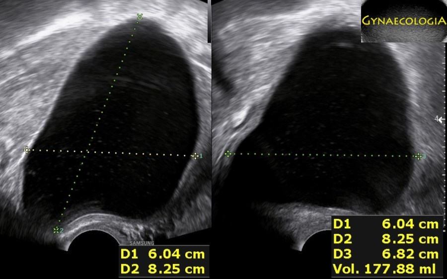
An asymptomatic 54-year-old patient was sent to us because of a “postmenopausal cyst”. She had a history of two vaginal deliveries and no abdominal surgeries. Our sonographic evaluation demonstrated a 8.3×6.8×6.0cm anechoic cyst on the posterior uterine surface and a normal right ovary. The left ovary could not be demonstrated on ultrasound. We suggested laparoscopic excision of the cyst.
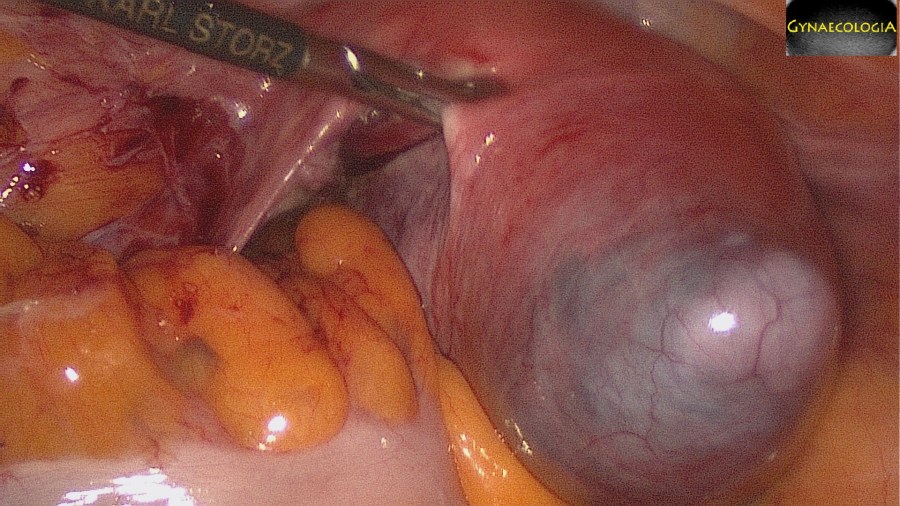
Intraoperatively we found that the left adnexa was missing. Adnexal agenesis has a rare occurence. Furthermore there was a large cyst on the posterior uterine wall, which could be succesfully excised. The cyst contents were clear. The uterine surface required suturing (3x figure of 8 stitches with PDS 0). The postoperative histology confirmed a benign, mesothelial cyst of the uterus.
Mesothelial cysts appear as single or multiple, thin-walled inclusion cysts derived from benign mesothelioma. Such cysts can occur at any abdominal peritoneal surface, commonly the round ligament, mesentery, and peritoneum. Uterine mesothelial cysts are extremely rare. Surgical excision offers definitive treatment.
Or click here to watch the full video on youtube.
Bibliography
- Mo, S. P., Wang, M. Y., & Li, J. K. (2019). Mesothelial cyst of uterine corpus misdiagnosed as leiomyoma. Chinese medical journal, 132(12), 1502–1503. https://doi.org/10.1097/CM9.0000000000000291
