

Borderline ovarian tumors (BOT) are epithelial tumors with:
- slow growth rate and
- low invasive potential (1).
Presence of vascularized and irregular papillary projections in a unilocular cystic mass in the absence of other abnormal findings should arise the suspicion of a borderline ovarian tumor (1, 2, 3). The presence of multiple septa may hint towards a mucinous borderline ovarian tumor (1).
If the solid component of the cyst demonstrates an acoustic shadow, this isn’t likely to be a borderline ovarian tumor and could be a cystadenofibroma (4).
Instead, the presence of thin-walled, microcystic formations along the borders of the solid component (typically 1-3mm in size) is thought to be pathognomonic of borderline ovarian tumors (5).
The presence of bilateral, irregular, vascularized solid masses with papillary architecture and internal branching which encase normal-appearing ovaries should direct the diagnosis towards the serous surface papillary subtype of borderline tumors (6).
We finally suggest using the Assessment of Different NEoplasias in the adneXa (ADNEX) model created by the International Ovarian Tumor Analysis (IOTA) group as it has been shown to be very accurate in the preoperative diagnosis of ovarian tumors by calculating multiple imaging characteristics (7).
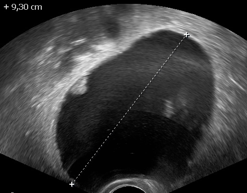
Irregular papillary projection in a unilocular cyst. 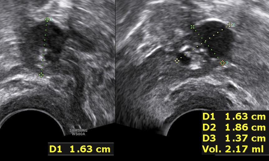
A small borderline tumor with presence of microcystic formations. 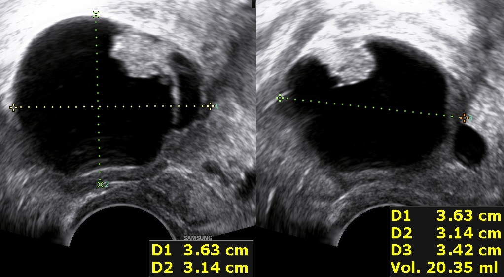
Another borderline tumor with microcystic formations. 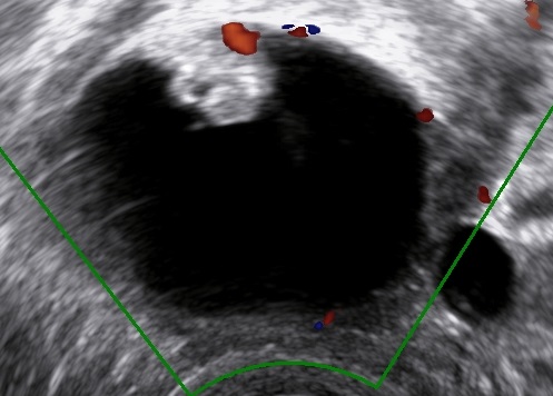
Vascularized, irregular papillary projection.
Bibliography
- Exacoustos, C., Romanini, M.E., Rinaldo, D., Amoroso, C., Szabolcs, B., Zupi, E. and Arduini, D. (2005), Preoperative sonographic features of borderline ovarian tumors. Ultrasound Obstet Gynecol, 25: 50-59. doi:10.1002/uog.1823
- Pascual, M. A., Tresserra, F., Grases, P. J., Labastida, R., & Dexeus, S. (2002). Borderline cystic tumors of the ovary: gray-scale and color Doppler sonographic findings. Journal of Clinical Ultrasound : JCU, 30(2), 76—82. https://doi.org/10.1002/jcu.10028
- Franchi, D., Boveri, S., Fruscio, R., Fischerova, D., Guerriero, S., Moruzzi, M.C., Colombo, N., Timmerman, D., Valentin, L. and Testa, A.C. (2013), Imaging in gynecological disease (8): ultrasound characteristics of recurrent borderline ovarian tumors. Ultrasound Obstet Gynecol, 41: 452-458. doi:10.1002/uog.12276
- Timor‐Tritsch, I.E., Yoon, E., Monteagudo, A., Ciaffarano, J., Brandon, C., Mittal, K.R., Wallach, R.C. and Boyd, L.R. (2019), Ultrasound and Histopathologic Correlation of Ovarian Cystadenofibromas: Diagnostic Value of the “Shadow Sign”. J Ultrasound Med, 38: 2973-2978. doi:10.1002/jum.15003
- Timor‐Tritsch, I.E., Foley, C.E., Brandon, C., Yoon, E., Ciaffarrano, J., Monteagudo, A., Mittal, K. and Boyd, L. (2019), New sonographic marker of borderline ovarian tumor: microcystic pattern of papillae and solid components. Ultrasound Obstet Gynecol, 54: 395-402. doi:10.1002/uog.20283
- Park, S. B., Kim, M. J., Lee, K. H., & Ko, Y. (2018). Ovarian serous surface papillary borderline tumor: characteristic imaging features with clinicopathological correlation. The British journal of radiology, 91(1088), 20170689. https://doi.org/10.1259/bjr.20170689
- Van Calster, B., Van Hoorde, K., Froyman, W., Kaijser, J., Wynants, L., Landolfo, C., Anthoulakis, C., Vergote, I., Bourne, T., & Timmerman, D. (2015). Practical guidance for applying the ADNEX model from the IOTA group to discriminate between different subtypes of adnexal tumors. Facts, views & vision in ObGyn, 7(1), 32–41.
