

Definition
A smooth muscle tumour of the uterine body, the visceral adnexa, or the uterine ligaments, which bears features that preclude an unequivocal diagnosis of leiomyosarcoma, but that do not fulfill the criteria for leiomyoma or its variants, and raises concern that the neoplasm may behave in a malignant fashion (Kurman, R. J., 2014, Patel, V., 2020).
Synonym: Atypical smooth muscle neoplasm (Kurman, R. J., 2014).
Epidemiology
There appears to be a universal agreement that the condition is rare, however exact data is lacking, possibly due to varying pathologic diagnostic criteria. Patient age at presentation appears similar to myomas (Ip, P. P., et al, 2010, Zheng, Y. Y. et al, 2020).
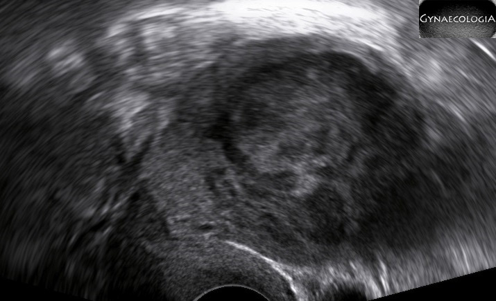
Clinical Image
Similar to myomas:
- Abnormal vaginal bleeding.
- Anemia.
- Pelvic mass.
- Pressure symptoms (Ip, P. P., et al, 2010, Oh, J., et al, 2019).
Diagnostics
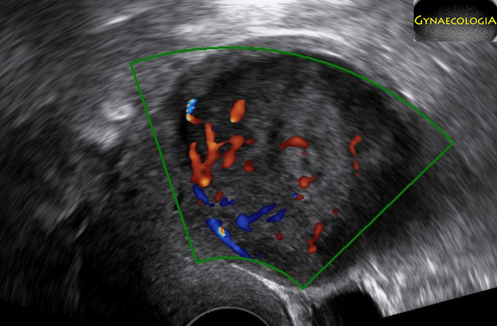
Ultrasound and MRI have been used extensively in an effort to distinguish among leiomyomata, leiomyosarcomata and even STUMP. Understanding the pathology behind these conditions, it shouldn’t come as a surprise that there is difficulty in setting pathognomonic features for each one (Oh, J., et al, 2019).
The following chart may be helpful when trying to establish a differential diagnosis:
| Leiomyoma | STUMP | Sarcoma | |
|---|---|---|---|
| Size | Variable | Variable | Usually already large when found |
| Borders | Regular | Regular | Could be irregular |
| Echogenicity | Isoechogenic or mixed | Isoechogenic or mixed | Inhomogenous |
| Calcifications | May appear | Absent | |
| Acoustic Shadows | Variable | Absent | Absent |
| Gynecological symptoms | May appear | Usually yes | Usually yes |
| Anechogenic areas | May appear | Yes | Yes, irregular |
| Vascularization | Circumferential and may even be intralesional | Circumferential and intralesional | Circumferential and intralesional |
| (Woźniak, A., & Woźniak, S., 2017) | (Cotrino, I., et al, 2020) | (Ludovisi, M., et al, 2019) |
Management
Total hysterectomy provides the definite surgical treatment in cases of STUMP (Zheng, Y. Y., et al., 2020).
Oncological follow-up is suggested.
- first three years -> every three months
- 4th and 5th year -> every six months
- after 5 years -> annual surveillance
STUMPs may recur as STUMPs or as leiomyosarcomas.
* If the diagnosis of a STUMP arises after a myomectomy where the tumor has been sufficiently excised, fertility preservation is possible, however it is imperative that both the gynecologist and the patient should remain vigilant for the danger of recurrence (Ip, P. P., et al, 2010).
Prognosis
After adequate treatment prognosis may be excellent. (Oh, J., et al, 2019). Recurrence is a threat, but it only seems to occur in uterine preservation surgeries (Zheng, Y. Y., et al., 2020).
Bibliography
- Oh, J., Park, S. B., Park, H. J., & Lee, E. S. (2019). Ultrasound Features of Uterine Sarcomas. Ultrasound quarterly, 35(4), 376–384. https://doi.org/10.1097/RUQ.0000000000000454
- Zheng, Y. Y., Liu, X. B., Mao, Y. Y., & Lin, M. H. (2020). Smooth muscle tumor of uncertain malignant potential (STUMP): a clinicopathologic analysis of 26 cases. International journal of clinical and experimental pathology, 13(4), 818–826.
- Kurman, R. J. (2014). Who classification of tumours of female reproductive organs. International Agency for Research on Cancer.
- Patel, V., Xing, D., Feely, M., & Schoolmeester, J. K. (2020). Smooth Muscle Tumors of the Visceral Adnexal and Uterine Ligaments and Adnexal Connective Tissue: A Clinicopathologic Study of 67 Cases. International journal of gynecological pathology : official journal of the International Society of Gynecological Pathologists, 39(1), 55–67. https://doi.org/10.1097/PGP.0000000000000578
- Ip, P. P., Tse, K. Y., & Tam, K. F. (2010). Uterine smooth muscle tumors other than the ordinary leiomyomas and leiomyosarcomas: a review of selected variants with emphasis on recent advances and unusual morphology that may cause concern for malignancy. Advances in anatomic pathology, 17(2), 91–112. https://doi.org/10.1097/PAP.0b013e3181cfb901
- Cotrino, I., Carosso, A., Macchi, C., Baima Poma, C., Cosma, S., Ribotta, M., Viora, E., Sciarrone, A., Borella, F., & Zola, P. (2020). Ultrasound and clinical characteristics of uterine smooth muscle tumors of uncertain malignant potential (STUMPs). European journal of obstetrics, gynecology, and reproductive biology, 251, 167–172. https://doi.org/10.1016/j.ejogrb.2020.05.040
- Ludovisi, M., Moro, F., Pasciuto, T., Di Noi, S., Giunchi, S., Savelli, L., Pascual, M.A., Sladkevicius, P., Alcazar, J.L., Franchi, D., Mancari, R., Moruzzi, M.C., Jurkovic, D., Chiappa, V., Guerriero, S., Exacoustos, C., Epstein, E., Frühauf, F., Fischerova, D., Fruscio, R., Ciccarone, F., Zannoni, G.F., Scambia, G., Valentin, L. and Testa, A.C. (2019), Imaging in gynecological disease (15): clinical and ultrasound characteristics of uterine sarcoma. Ultrasound Obstet Gynecol, 54: 676-687. https://doi.org/10.1002/uog.20270
- Woźniak, A., & Woźniak, S. (2017). Ultrasonography of uterine leiomyomas. Przeglad menopauzalny = Menopause review, 16(4), 113–117. https://doi.org/10.5114/pm.2017.72754
